Pathology resident persona
Integrating artificial intelligence to enhance pathologist workflows in identifying mitoses

UX Designer
January 2022 – April 2022
Hongyan Gu (Project Lead), Shirley Tang (UX Designer), 1 engineer
User Personas, User Flows, Wireframing, Prototyping, Usability Testing
Adobe XD, Miro
Access this project's publication, “Augmenting Pathologists with NaviPath: Design and Evaluation of a Human-AI Collaborative Navigation System“ at the ACM digital library.
Areas of mitosis can suggest the presence of meningioma, which is something pathologists routinely check for when evaluating digitized tissue scans. To leverage artificial intelligence in the medical space, we were challenged to find an opportunity to help pathologists save time and effort in the mitosis-identifying process without disrupting their current workflow.
Our solution is NaviPath: an AI-enabled navigational tool that helps pathologists identify areas of mitosis based on a set of interactive cellular criteria telling users where to look. NaviPath is built based on hierarchical AI recommendations, customizable recommendations based on multiple criteria, and cue based navigation.
Overall, we found that navigation efficiency, participant precision, and recall for identifying target pathology patterns were significantly improved (p<0.05) using NaviPath.
As a UX designer, I led the research and design process from synthesizing user research, designing a prototype, and moderating usability tests to uncover insights on how pathologists interact with AI systems.
I analyzed 6 user interviews that focused on learning about the pathologist navigation process.
We found that pathologists tended to:
1. View scans holistically at a low magnification, then at regions of interest in higher magnifications
2. Use macroscopic patterns visible in low magnifications to locate regions of interest
3. Search systematically in high magnifications
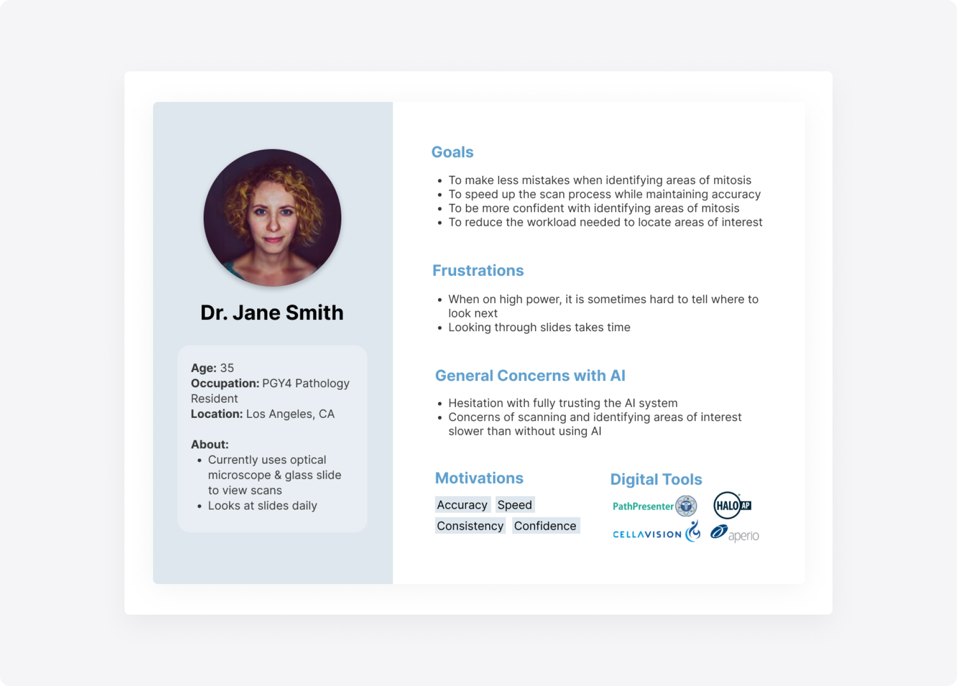
Using this data, I created a post-graduate year 4 pathology resident persona to guide our focus on the target users.
Pathology resident persona
Previous literature suggests pathologist constraints with time, focus, and price when using digital pathology tools compared to traditional microscopes. Additionally, pathologists have been found to get distracted when AI systems directly point out regions of interest.
While we wanted the system to integrate AI, it was important to encourage more human engagement, rather than leading with AI.

One of the technical needs involved four levels of magnification that pathologists could use to look for mitosis: a tissue level that shows the entire scan, a local level that magnifies enough to show macroscopic patterns, a high power field level that further magnifies and shows cells, and a cell level that is the highest magnification possible in the system.
I created a user flow to visualize how interactions between levels would take place, in which we found that the system would essentially be a tree traversal: users would be able to follow down a path from lowest to highest magnification, but would not be able to skip across levels.

User tasks and flows demonstrate interactions with the magnification levels
System design requirements shifted as we continued to uncover user needs and technical constraints. These requirements included the system being multi-faceted, self-explainable, and adjustable.
We also had to prioritize a couple of points across different dimensions:
1. Considering users: how do we logically present the recommendations to pathologists?
2. Considering human-computer interaction: what designs can properly convey our intended function?
3. Considering AI: what is needed for mitosis detection and how can we justify our recommendations?
Wireframes for the tissue and cell levels helped fuel exploration around how to incorporate potential elements of the system based on research. We iterated on prioritized features such as recommendation boxes and justification pop-ups.

Low-fidelity wireframes of the system interface and recommendation slider
One challenge was determining how to represent the criteria that users could toggle to actively filter recommendations. During the first round of testing, we opted to include six different criteria in the design based off research findings on characteristics that pathologists used while searching for mitosis.
By the end of six weeks, we began usability testing on an MVP with pathologists to determine the validity of our prototype.

Design iterations of the system interface
The goals of usability testing included:
1. Checking if users could understand and use the criteria provided by the system
2. Determining whether the assumptions about the users’ workflow correspond with the users’ mental models, and
3. Exploring whether there are any design inconsistencies or pain points within the user interface or interactions.
The session consisted of four parts: a project overview, system function section, co-design session, and design review.

Moderating usability testing sessions at the lab
Details pertaining to copy and UI such as the language of justifications and sizes of recommendation boxes on the screen were mentioned most frequently among participants.
Participants commented that the criterion of "cellular intensity" was least relevant when locating mitosis. It was also noted that the wording of "mitosis uncertainty" was confusing as opposed to "mitosis certainty".
Another finding was the preference for using heat maps to show mitosis probability and cellular density while evaluating the presence of mitosis, which was added into the next iteration.
We shifted our focus from testing ease of use to evaluating accuracy and effectiveness when using the system.
The next round of testing aimed at validating NaviPath against using only human or only AI systems. Research questions focused on asking:
1. Does NaviPath lead to higher quality mitosis reporting?
2. Will NaviPath save pathologists’ time and effort?
3. Does NaviPath add value to participants’ workflow?
Three testing conditions were created, where pathologists (n=15) either (1) navigated a pathology tumor scan viewer without any AI assistance, (2) navigated using NaviPath, or (3) were compared to an AI-automatic reporting system.
Participant interactions, reporting of mitoses, and time-elapsed were recorded.

NaviPath MVP displaying hotspots of mitosis areas
We found that NaviPath produced significantly better performance compared to manual navigation and higher precision and recall compared to the AI-only system.
Though participants spent more time on average using NaviPath, they saw significantly more mitoses in unit time compared to manual navigation.
And lastly, panning and zooming interactions were significantly reduced using NaviPath compared to manual navigation.

AI recommendations and flexibility in toggling various cellular criteria enhance the ability to find areas of mitoses in digitized tissue scans.

